The Medical Imaging Laboratory, which requires the techniques and technologies needed for radiological examinations in the diagnosis or treatment of diseases, has been put into service as a training and research laboratory.
The Medical Imaging Laboratory, which is used for educational purposes, has a well-equipped team to train students for patient approaches and positions to consolidate their knowledge before going on the field.
In addition to theoretical knowledge, students have the opportunity to work in a real laboratory environment with advanced practical and practical training, and they can take a step into their professional working lives with the experience they have gained when they graduate.


Medical Imaging Techniques Department Head
444 1 428 - 57003


| General Information | It aims to conduct screening by adjusting the angle and intensity of the area in which the X-ray will be applied. |
| Brand & Model | Kolimatör Aselsan D110C |
| Technical Details | Vibrations which has 3000 MHz frequency are created in the 10 diameter chambers. Such high frequency electromagnetic waves are transmitted to the channel in the middle of these chambers.
|
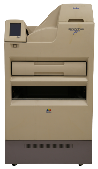
| General Information | The tape, transmitted by the stative after placing the x-ray film, is transferred to the imaging device. The device then scans the tape and transmits the image to the x-ray film. Thus, helps the writing and imaging of radiogram to the film. |
| Brand & Model | Konica Drypro 751 |
| Technical Details | Filtration and Half-Value Thickness Test X-Ray Field and Beam Field Suitability (Collimation) and Perpendicularity Test Focus Point Size and Separation Efficiency Test Automatic Beaming Control Test |
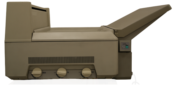
| General Information | X-ray’s feature of passing through tissue is the fundamental reason behind their usage in diagnostic radiology. Its fluorescence and photographic properties helps to obtain an image. Obtained film tapes are processed through CR film printing machine. The film obtained from the printer is delivered to the patient. |
| Brand & Model | Processor SRX-101A |
| Technical Details | Heat resistant tube. Vacuumed for longer use and efficient x-ray production. 5cm2 tube entrance Tube in cast for preventing unnecessary x-ray spreading. |
| General Information | X-ray’s feature of passing through tissue is the fundamental reason behind their usage in diagnostic radiology. Its fluorescence and photographic properties helps to obtain an image. Obtained film tapes are processed through CR film printing machine. The extent of the absorption of x-ray beams will differentiate, since the atoms and tissues forming the human body consist of different thickness, intensity and weight. X-ray beams forms an image by penetrating through tissue with different absorption and penet |
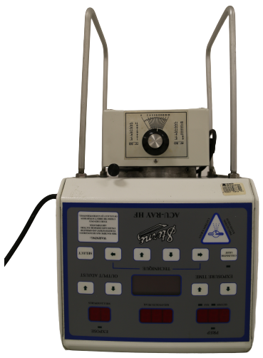
| Brand & Model | Turay HF 50-R |
| Technical Details | Computed Tomography (CT) |
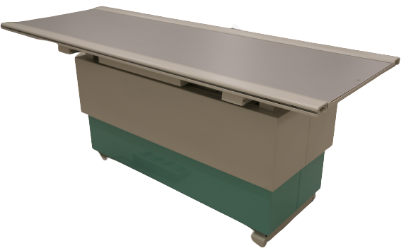
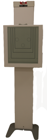
| General Information | Floating/elevator table is simple to use but an efficient equipment. |
| Brand & Model | Floating/Elevator Table |
| Technical Details | Can be moved to left or right. Rail system No electric current
|
*
| Laboratuvar | Cihaz Adı | Test Adı/Tedavi Parametreleri/Yapılan Uygulamalar |
|---|---|---|